Figure 1
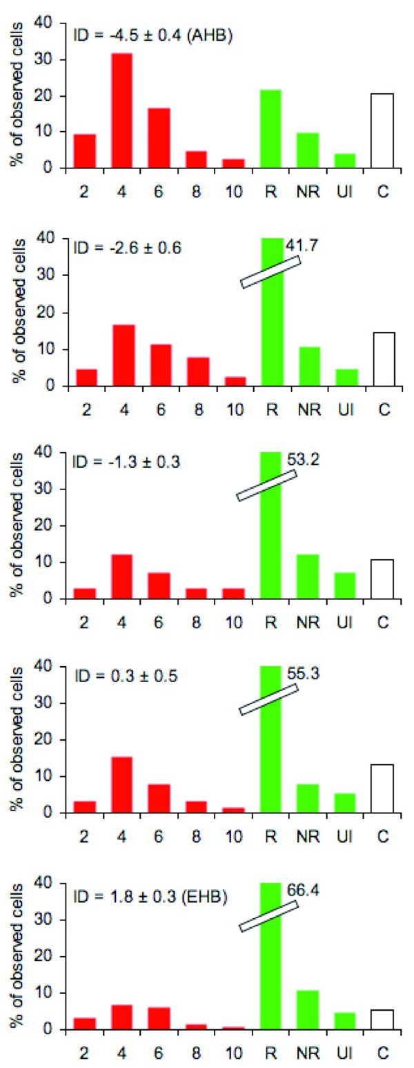
Removal of pupae artificially infested with a Varroa destructor mite in colonies with different Africanization levels. Selection of colonies. The level of Africanization (morphometric analysis) of 60 colonies of beekeepers apiaries was determined following the procedure described by Rinderer et al. (1993). Among them, 25 colonies were chosen for their homogeneous distribution along an Africanized-European cline, in groups of 5 colonies. The Africanization degree (ID) is the projection on the discriminant function 1 in the multivariate analysis performed by Rinderer et al. (1993) and varies from –6 (most Africanized colonies, top graph) to 4 (most European ones, bottom graph). Artificial infestation. 30 mites were collected from emerging brood (i.e. at a phase when they are able to reproduce) and were introduced into 30 cells containing pupae from the same colonies (to avoid detection for odor of another colony) that had been capped for less than 3 h. Additionally, 30 cells were uncapped and immediately recapped as controls. Determination of removal abilities. The cells containing artificially infested pupae were observed every 2 days over 10 days to determine if the bees removed the pupae. The 10th day, all remaining cells were uncapped to determine if they were still infested, and if so, if the mite inside had reproduced. The figure shows for each 5-colony group of colonies from most Africanized (top graph) to least Africanized (bottom graph), the percent of observed pupal cells in each of the following categories: cells uncapped in each 2-day period over 10 days after capping (« 2 » to « 10 »); cells in which mites reproduced normally (« R »); cells still capped, but in which mites did not reproduce (« NR »); cells no longer infested (« NI »); control cells uncapped in the first 10 days after capping (« C »). Overall, the more Africanized (i.e. unselected) the colonies, the more they detected pupae containing reproducing Varroa mites.