Free Access
Figure 3

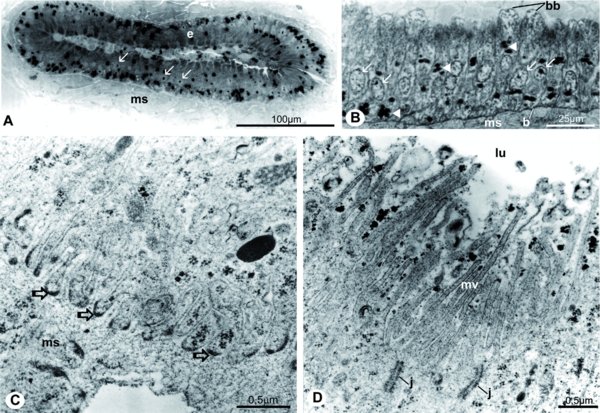
(A–B) Histology of adult seminal vesicles: (A) just-emerged, with reduced lumen, (B) epithelial detail of a one day-old adult with bubble-like projections (bb), (C–D) TEM of the seminal vesicle of one day old adult; (C) basal cellular portion, note anchorage structures (open arrows) in cellular infoldings, (D) apical cell portion. (b) basal layer, (e) seminal vesicle epithelium, (j) cell junctions, (lu) seminal vesicle lumen, (ms) muscular layer, (mv) microvilli, (arrow) epithelial cell nuclei, (arrowhead) dark lipid droplets.


